
Patients diagnosed with early rectal neoplasms or complex polyps frequently explore treatment pathways that maintain normal bowel anatomy and avoid conventional abdominal incisions. Transanal Minimally Invasive Surgery (TAMIS) is an established endoluminal approach designed for organ-preserving local excision. By operating through the natural transanal corridor, colorectal surgeons can remove selected benign rectal lesions and, in carefully selected cases, early-stage rectal cancers.
Colorectal conditions require rigorous diagnostic workups, dedicated endoluminal platforms, and individualized patient selection. Dr. Daniel Serralta, Consultant colorectal surgeon and proctologist, provides minimally invasive laparoscopic and transanal surgical evaluations in Dubai.
What is TAMIS Surgery?
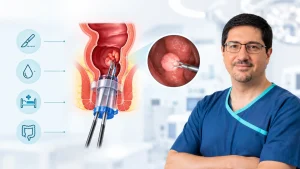
Transanal Minimally Invasive Surgery (TAMIS) represents an advanced surgical technique within proctology and colorectal surgery. Traditionally, accessing lesions situated in the middle to upper rectum required major open or laparoscopic abdominal operations. The introduction of TAMIS has expanded options for local excision by combining flexible single-port transanal access devices with standard laparoscopic instruments and high-definition optical systems.
During a TAMIS procedure:
- A specialized multichannel transanal access port is positioned within the anal canal.
- The rectal cavity is gently insufflated with medical carbon dioxide (CO₂) to create a stable, pressurized viewing space (pneumorectum).
- Using high-definition laparoscopic cameras and fine articulating instruments, the surgeon visualizes the mucosal surface and excises the targeted lesion with clear surgical margins.
Because access is achieved entirely through the natural anatomical opening, patients avoid external abdominal incisions, abdominal wall muscle trauma, and incisional wound complications.
Clinical Indications and Candidate Selection
Careful patient selection based on pre-operative clinical and radiological staging is critical for achieving appropriate oncological outcomes. Not all rectal lesions are suitable for local excision.
A comprehensive pre-operative evaluation includes:
- High-resolution pelvic MRI: Evaluates depth of wall penetration and screens for regional lymph node involvement.
- Endorectal Ultrasound (ERUS): Assesses mucosal and submucosal layer differentiation.
- Full Colonoscopy with Histopathology: Evaluates the entire colon for synchronous lesions and provides initial tissue diagnosis.
Primary Indications:
- Large Benign Adenomas: Extensive or flat polyps not amenable to standard endoscopic mucosal resection (EMR) or endoscopic submucosal dissection (ESD).
- Selected Early-Stage Malignancies (cT1N0): Low-risk, well-to-moderately differentiated invasive adenocarcinomas without lymphovascular invasion or adverse histological features.
- Rectal Neuroendocrine Tumours (NETs): Localized, well-differentiated carcinoid tumors typically measuring usually under 2 cm.
- High-Grade Dysplastic Lesions: Mucosal abnormalities carrying significant risk of malignant progression if not excised.
- Palliative Interventions: Symptom relief (e.g., local bleeding or partial obstruction) in patients medically unfit for major abdominal resection.
Surgical Protocol: Step-by-Step
The TAMIS procedure follows a standardized endoluminal protocol under general anesthesia:
- Preparation: Patients complete standard bowel preparation before surgery to ensure clear endoluminal visualization.
- Positioning: The patient is placed in either lithotomy or prone jackknife position, tailored to the anterior, posterior, or lateral orientation of the rectal lesion.
- Port Placement & Insufflation: The lubricated transanal access device is placed in the anal canal and linked to a continuous-pressure CO₂ insufflator.
- Circumferential Demarcation: The perimeter of the lesion is marked using cautery dots to maintain targeted histological margins.
- Excision: A full-thickness or partial-thickness excision is performed through the submucosa and muscular layer down to perirectal fat, as clinically indicated.
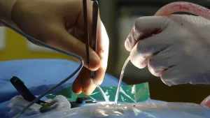
- Defect Closure: The excised specimen is oriented for histopathological review. The rectal wall defect is irrigated and closed transversely using absorbable barbed sutures to prevent lumen narrowing and minimize delayed postoperative bleeding.
Clinical Benefits of Transanal Excision
The primary advantage of transanal local excision lies in the complete absence of external abdominal incisions. By avoiding entry through the abdominal wall, because TAMIS does not require an abdominal incision, it reduces the risk of incision-related complications such as an abdominal wound infection or incisional hernia. In addition, because the surgical dissection is confined strictly to the rectal wall, TAMIS avoids the extensive pelvic dissection used in some radical operations, but urinary, sexual and bowel function outcomes depend on the procedure and individual clinical factors. Furthermore, modern flexible single-port systems overcome the anatomical constraints of traditional rigid transanal retractors, offering surgeons the maneuverability and optical reach needed to evaluate and excise lesions situated in the middle and upper rectum.
Potential Risks and Clinical Considerations
Although transanal minimally invasive surgery is an organ-preserving, low-impact intervention, it entails specific procedural considerations that must be evaluated prior to surgery. For lesions located along the upper anterior rectal wall above the peritoneal reflection, full-thickness excision carries a potential risk of entering the peritoneal cavity, which may necessitate immediate laparoscopic closure. Patients may also experience delayed bleeding from the internal suture line, occasionally requiring clinical observation or localized endoscopic intervention.
Minor, transient alterations in bowel patterns—including mild urgency, occasional soiling, or variable stool frequency—can occur during the initial healing phase as mucosal inflammation resolves. Finally, if the definitive histopathological evaluation reveals involved margins, high-risk biological features, or deeper muscular invasion (such as T2 disease), completion radical surgery may be indicated to ensure adequate oncological disease control.
Recovery Timeline and Aftercare
Postoperative recovery following a transanal procedure progresses rapidly compared to traditional open or laparoscopic resections. Some patients can be discharged the same day or within 24 hours, depending on their recovery, clinical findings and the treating team’s assessment. Discomfort is typically mild to moderate and well controlled using standard oral analgesics rather than heavy opioid regimens. Clear fluids and light meals can be resumed shortly after waking from anesthesia.
To shield the endoluminal suture line from unnecessary stress, patients are advised to maintain consistent hydration, follow a fiber-rich diet, and take gentle stool softeners to prevent straining during defecation. Routine sedentary activities and Light activity can often be resumed gradually, following the surgeon’s postoperative instructions. Although heavy lifting and vigorous exercise should be deferred for generally two to three weeks to allow internal tissue consolidation. A scheduled postoperative review is then conducted to examine tissue healing and formulate any subsequent surveillance plans based on the final pathology report.
Consultation and Clinical Assessment in Dubai
For individuals diagnosed with rectal polyps or early-stage rectal lesions, specialized clinical assessment confirms whether transanal minimally invasive surgery is an appropriate option.
Dr. Daniel Serralta conducts diagnostic evaluations and surgical planning at Saudi German Hospital, Dubai.
Request a Consultation
If you have received an abnormal colonoscopy finding or have symptoms such as rectal bleeding or altered bowel patterns, a specialized colorectal evaluation is recommended.
To discuss diagnostic findings and explore whether TAMIS is appropriate for your case, schedule a clinical consultation with Dr. Daniel Serralta in Dubai.
Frequently Asked Questions:
1. What types of rectal lesions are suitable for TAMIS?
TAMIS is primarily suited for large benign rectal adenomas, localized neuroendocrine tumors, and selected early-stage (cT1N0) adenocarcinomas with favorable histological features confirmed on pre-operative imaging.
2. How long does recovery take after TAMIS?
Most patients return to light daily routines generally within 3 to 5 days. Healing time varies depending on the extent of excision and individual recovery. Follow-up helps assess healing and guide activity restrictions.
3. Can TAMIS affect bowel continence?
Because TAMIS avoids extensive sphincter disruption as a small number of patients experience temporary or, rarely, longer-term changes in continence. It generally preserves the rectal reservoir, normal baseline continence is typically maintained. Some individuals may experience mild, transient urgency or altered stool frequency while the internal rectal wall heals.
4. What type of anesthesia is required?
TAMIS is usually performed under general anesthesia; the anesthetist advises on the best option to ensure complete pelvic muscle relaxation, airway safety, and an uninterrupted operational field.
5. Is TAMIS always an alternative to major rectal surgery?
Not in all instances. TAMIS is an alternative only for carefully selected, early-stage, or benign lesions. If final pathology shows deeper tumor invasion (T2 or greater), lymph node involvement, or unfavorable biological features, secondary radical surgery may still be recommended.